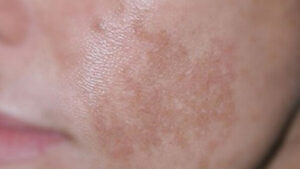
SKIN BLEMISH (MELASMA) TREATMENT AND PROTECTION FROM BLEMISHESMelasma is the formation of black or brown spots on the skin. Melasma is primarily characterized by discoloration or hyperpigmentation of the facial skin. Melasma usually appears along the central facial line, predominantly on the forehead, cheekbones, chin, and upper lip, and rarely on the neck and forearms.
Genetic predisposition, ultraviolet exposure, pregnancy, hormone therapy, cosmetics, and medications are the etiological factors that cause melasma. Although it is asymptomatic, the cosmetic disturbance it causes leads to psychological stress. Primary treatment options include topical hypopigmenting agents, chemical peels, laser therapy, and dermabrasion.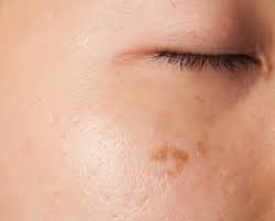 Although melasma can be seen at any age, race, and sex, it is more frequently observed between the ages of 30-55, particularly in Asian, Hispanic, and Black women with Fitzpatrick skin phototypes IV-VI (categorized into 6 groups from very fair (skin type I) to very dark (skin type VI) according to the Fitzpatrick skin type scale) who are exposed to high actinic radiation. 90% of patients are women. Its true prevalence is unknown. Its incidence during pregnancy is 50-70%, and 5-34% among those using oral contraceptives (OCPs). The frequency of melasma increases during summer months and decreases in winter.
The exact cause of melasma is not fully known. Genetic predisposition and ultraviolet (UV) exposure, conditions with elevated estrogen levels (pregnancy, use of oral contraceptives (OCPs), hormone replacement therapy), cosmetics, photosensitizing medications, autoimmune thyroid disorders, race, psychosomatic factors, hepatic dysfunction, parasites, and nutritional deficiencies can cause melasma. There are also melasma patients who have no association with any of these factors. It is reported that melasma falls into the idiopathic category in at least one-third of female patients and in the majority of male patients. Although the role of genetic factors is not fully understood, a family history is present in 50% of cases. Sunlight has long been known to be one of the primary factors in the development of melasma. The higher prevalence in geographic regions with intense sunlight, its increase in summer months, its development on sun-exposed areas of the body, and the frequent history of sunlight exposure at the onset of lesions suggest that this factor triggers the development of melasma, especially in individuals with genetic and hormonal predispositions. Melasma appearing on the face, which is considered physiological during pregnancy, is also referred to as the mask of pregnancy or chloasma. It is observed in 50-70% of pregnant women. This condition, which is progressive throughout pregnancy, increases with UV exposure. Although it resolves within 1-5 years postpartum in most cases, it can sometimes be persistent. It may recur or worsen in subsequent pregnancies or with OCP use.
In the etiopathogenesis of melasma, numerous photosensitizing and phototoxic drug classes are held responsible, most notably hormone-containing OCPs, anticonvulsants (phenytoin), phenothiazines (chlorpromazine), tricyclic antidepressants, antiarrhythmics (amiodarone), antimalarials (chloroquine, hydroxychloroquine, quinacrine, and mefloquine), antibiotics (tetracycline and minocycline), and heavy metals (gold and silver salts, iron, bismuth). Clofazimine, bleomycin, busulfan, rifampin, dapsone, and zidovudine are other drugs that rarely cause melasma-like hyperpigmentation. Additionally, cases of melasma that developed with isotretinoin and spironolactone treatments and regressed upon discontinuation have also been reported. Drug-induced melasma generally resolves within months after stopping the medication. Cosmetics cause melasma through phototoxic mechanisms. Perfumed and colored cosmetic products containing bergamot oil or furocoumarins, scented soaps, and facial cleansers can cause facial hyperpigmentation. Heat applications are also recognized to play a role in the etiology of melasma. Melasma can appear on the upper lip mustache area following hot wax application. This is frequently reported by patients. Studies have shown that heat application in cell cultures leads to elongation of melanocyte dendrites, an increase in cell size, and an increase in tyrosinase activity, similar to UVB exposure.
Although melasma lesions are predominantly located on sun-exposed areas of the face (98.3%), they may rarely be seen in different areas such as the neck, forearms, trunk, nipples, and genital region. Lesions may be linear or guttate. They typically present as solitary or, more commonly, symmetrically distributed multiple macules. They are characterized by well-demarcated, irregular light-to-dark brown or gray macules and patches. Although the color is generally homogeneous, it can sometimes display a mottled appearance. Mucosal involvement is not observed. Three clinical patterns have been described based on lesion distribution: centrofacial, malar, and mandibular:
1. Centrofacial pattern: The most common distribution pattern, seen in 63% of patients. In this pattern, lesions are located on the forehead, cheeks, nose, upper lip, and chin (mask-like).
2. Malar pattern: A clinical pattern where lesions are localized to the cheeks and the bridge of the nose (butterfly-like). It is seen in 21% of patients.
3. Mandibular pattern: Lesions are located on the rami of the mandible. It is the least common clinical pattern of melasma, observed in 16% of patients.
Histopathological examinations show no difference between clinical patterns. Epidermal-type melasma clinically appears light brown. Dermal-type lesions are clinically blue or ash-gray in color. Mixed-type lesions are generally light brown clinically.
The diagnosis of melasma is generally made through medical history and skin examination, while the histological localization of lesions is determined using Wood’s lamp examination. Patients should be evaluated regarding the onset and duration of hyperpigmentation, aggravating and relieving factors, family history, and the use of medications and cosmetics. Patients are evaluated under natural light for lesion distribution, homogeneity, extent, and pigmentation density. Wood’s lamp helps determine the depth within the skin layers at which the pigmentation disorder occurs. The filter in this lamp blocks all rays outside the 320-400 nm band. Because the filtered light emitted from this lamp has a shorter wavelength than visible light, it easily highlights epidermal pigmentation changes. Since it does not penetrate well into the deeper layers of the dermis, reflection is low for dermal changes. As a result of this differential reflection characteristic, melasma is categorized into three distinct types under Wood’s lamp examination: epidermal, dermal, and mixed. In the epidermal type, hyperpigmented macules appear sharply demarcated and darker brown under Wood’s lamp examination. In the dermal type, lesions that appear blue-gray in natural sunlight appear faint, less distinct, and with irregular margins when examined under Wood’s light. In the mixed type, because both dermal and epidermal melanin are present, a brown-gray mottled appearance is observed with darkening in some areas and fading in others. Wood’s lamp examination is crucial for treatment selection and determining prognosis, as the treatment response of patients with epidermal-type melasma is significantly better compared to the dermal type.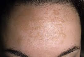 Berloque dermatitis, a pigmentation disorder typically developing on the sides of the neck and behind the ears, characterized by asymmetrical macules and patches and caused by a phototoxic reaction to furocoumarins in perfumes and colognes, is differentiated from melasma by patient history and lesion localization. Likewise, any inflammatory condition developing at the dermoepidermal junction can lead to post-inflammatory hyperpigmentation. It is distinguished from melasma by a history of eruption prior to pigmentation and by histopathological examination.
Untreated lesions in melasma are persistent and do not resolve spontaneously. The disease progresses slowly over time. Complete remission is rare, and recurrence following treatment is common. Rarely, spontaneous remission may be observed with the cessation of sun exposure.
Since melasma is a chronic disease, its response to treatment is poor. It generally requires a long-term treatment period. In general, epidermal and mixed-type melasma respond better to treatment than dermal melasma. Melasma treatment is particularly challenging in dark-skinned patients and is limited by the adverse effects of therapies. Post-treatment recurrence is also a significant issue. Treatment is not recommended during pregnancy, lactation, and in women using OCPs.
Melasma is a distressing condition for most patients from a cosmetic standpoint. Therefore, when selecting therapeutic agents, those that are locally acting and safe in the long term should be preferred. The goal of treatment is to achieve an acceptable cosmetic appearance and maintain it over the long term.
The principles of melasma treatment are primarily categorized under five main headings:
1. Sun protection
2. Suppression of melanocyte activity
3. Suppression of melanin synthesis
4. Removal of existing melanin pigment
5. Degradation of melanin granules in the tissue
Sun protection; for successful treatment, protecting the patient from the sun and providing patient education are essential. Since sunscreen products are known not to effectively block all ultraviolet wavelengths, other physical methods such as sun avoidance, protective clothing, hats, and window screens/protectors should also be utilized. In melasma patients, opaque, non-comedogenic, oil-free sunscreen products with an SPF of >15 for UVB and >30 for UVA should be preferred. Systemically administered chloroquine provides protection against UVA; indomethacin, vitamins C and E, and green tea against UVB; β-carotene against UVA and visible light; and fish oil against both UVA and UVB.
Suppression of melanocyte activity; since sunlight, pregnancy, birth control pills, fragranced cosmetics, and phototoxic drugs activate melanocytes, these must be avoided. Suppression of melanin synthesis; topical tyrosinase inhibitors—hydroquinone, a chemical molecule with a hydroxyphenol structure, inhibits tyrosinase in melanocytes, thereby preventing the conversion of tyrosine to dopa and thus inhibiting melanin synthesis.Hydroquinone;It is one of the most frequently used and most effective agents in the treatment of melasma. It can be applied alone or in combination with other therapeutic agents. In the treatment of melasma, it is used at concentrations of 2–10%, twice daily. Although 2% and 4% concentrations are available in our country, the safest concentration used is 2%. While 4% concentrations of hydroquinone are more effective, their side effects are greater. High concentrations of 8–10% are generally used in resistant melasma and for depigmentation in dark-skinned individuals. Its effect begins after the 4th week of treatment, optimal efficacy appears within weeks 6 to 10, and its use as a single agent requires a long treatment duration. It is particularly effective in epidermal-type melasma. Hydroquinone can be combined with agents such as sunscreens, antioxidants, tretinoin, glycolic acid, topical corticosteroids, kojic acid, and azelaic acid. Among combination therapies, the most popular is the “Kligman formula,” first described in 1975 by Kligman and Willis, containing 5% hydroquinone, 0.1% tretinoin, and 0.1% dexamethasone in a hydrophilic ointment. For this purpose, 0.01% fluocinolone acetonide in triple therapy was found to be more successful and had fewer side effects. This combination remains one of the most effective treatment modalities for melasma.
The side effects of hydroquinone in chronic use include contact dermatitis, permanent leukoderma, whitening of the surrounding skin, cataracts, colloid milium, pigmentation of the sclera and nails (pseudo-yellow nail syndrome), decreased skin elasticity, impaired wound healing, and a fish-like odor sensation. In some patients, exogenous ochronosis may be seen in sun-exposed areas due to the accumulation of homogentisic acid in the dermis. Although this condition can rarely be seen even with short-term use of 1–2% hydroquinone, it generally occurs when used frequently and at high concentrations. Therefore, long-term and high-dose use of hydroquinone should be avoided, and the use of hydroquinone at a maximum concentration of 2% should be recommended for maintenance therapy or during the summer months.
Endogenous ochronosis has been most frequently reported from South Africa and, to a lesser extent, from the United States. Although hydroquinone has been shown to cause DNA damage in animal studies, its carcinogenic potential is not fully known because no skin cancer or internal malignancy associated with topical use of the drug has been reported. Due to its side effects and potent efficacy, hydroquinone is not included in cosmeceutical products.Azelaic acid (AZA);AZA is a naturally occurring, non-toxic, 9-carbon dicarboxylic acid formed as a result of the oxidation of oleic acid. Pityrosporum species oxidize unsaturated fatty acids, leading to the production of AZA. It exerts antiproliferative and cytotoxic effects by inhibiting mitochondrial oxidoreductase activity and DNA synthesis. It is also a reversible in vitro competitive tyrosinase inhibitor. AZA is selectively toxic to hyperactive and abnormally proliferating melanocytes, but AZA has no toxic effect on normal melanocytes. In the treatment of melasma, it is generally used at a 20% concentration twice daily for a duration of 4–6 months. As monotherapy, it is effective in 60–90% of cases. More effective results are reported when combined with retinoic acid and topical steroids. AZA is generally well tolerated. Its most common side effect is local irritation at a rate of 5–10%, and tolerance develops over time. AZA is recognized as a good treatment option in patients who cannot tolerate hydroquinone therapy.Kojic acid;It is an antibiotic produced by fungi such as Aspergillus and Penicillium. It inhibits tyrosinase activity by chelating copper ions, thereby providing skin lightening. It is used at a concentration of 2–4% twice daily until cosmetic improvement is achieved. Its efficacy is virtually identical to that of 2% hydroquinone. Excellent response is obtained in combination with topical steroids and tretinoin. It can also be combined with hydroquinone and glycolic acid. The most significant side effect of kojic acid is the development of contact dermatitis.Tretinoin;Tretinoin is a vitamin A derivative, and its exact mechanism of action in melasma is not fully understood. It is thought to act by inhibiting tyrosinase and dopachrome conversion factor, thereby preventing melanin synthesis; reducing keratinocyte cohesion (keratolytic effect), blocking the transfer of melanin to keratinocytes, and dispersing melanin granules within keratinocytes. Tretinoin is effective in epidermal melasma at concentrations of 0.05-0.1%, but has no effect on dermal melasma. Although tretinoin can be effective alone in melasma, it can also be used in combination with other agents. It is applied once daily, at night only. Along with this, as with all other therapeutic agents, broad-spectrum sunscreens should also be used. It should not be used during the summer months. Its most significant disadvantage is the delayed onset of its effect and the high incidence of side effects. The most common side effect is retinoid dermatitis. It consists of burning, itching, erythema, and desquamation; as treatment continues, tolerance develops and these side effects decrease. Moisturizers and 1% hydrocortisone may be used to reduce side effects. Occasionally, post-inflammatory hyperpigmentation may develop following severe retinoid dermatitis.Arbutin;Arbutin is a glucopyranoside containing a hydroquinone molecule linked to glucose. It is among the most widely used skin-lightening agents worldwide. It is obtained from the dried leaves of plants such as bearberry, blueberry, and lingonberry. Its depigmenting effect is thought to result from the reversible inhibition of melanosomal tyrosinase activity and the inhibition of melanosome maturation. A 7% concentration is used in melasma. High doses may lead to paradoxical hyperpigmentation. Its synthetic forms, alpha-arbutin and deoxyarbutin, inhibit tyrosinase more potently. Although it is widely found in cosmeceuticals, there is insufficient literature data from studies regarding its efficacy in the treatment of PIH.Licorice root extract;Glabridin, found in licorice root extract at a concentration of 10% to 40%, suppresses tyrosinase activity; when applied at a concentration of 0.1% three times daily for one month, it provides a significant lightening in melasma color. It is present especially in certain cosmetic products at concentrations of 1% or higher. Its efficacy is 16 times greater than that of hydroquinone. Its clinically effective concentration remains unknown. It also exhibits an anti-inflammatory effect.Local Melanin Synthesis Inhibitors;
Ascorbic acid (Vitamin C);Ascorbic acid inhibits tyrosinase activity by enzymatically reducing o-quinone production, thereby preventing melanin synthesis. At the same time, through its antioxidant effect, it prevents the formation of free radicals that trigger melanogenesis and reduces UV absorption. It can be used alone or in combination therapies in the treatment of melasma. It has no side effects with topical use.Vitamin E (α-Tocopherol);Studies investigating the efficacy of α-tocopherol ferulate (a compound of α-tocopherol and ferulic acid) on melanogenesis in cultured human melanocyte cells have shown that this substance indirectly inhibits tyrosine hydroxylase activity. Researchers have suggested that tocopherol derivatives are more potent melanin formation inhibitors than arbutin and kojic acid, and that α-tocopherol ferulate acts by inhibiting the tyrosinase enzyme in addition to antioxidant mechanisms to prevent or treat UV-induced facial hyperpigmentation.Glutathione;It inhibits eumelanin synthesis by increasing the formation of pheomelanin from dopaquinone.Non-Selective Melanogenesis Suppression
Indomethacin;A 5% concentration of indomethacin exhibits a corticosteroid-like effect. It is particularly effective in epidermal-type melasma located in the vermilion region. It also has a sun protection effect equivalent to SPF 3.Topical steroids;The mechanism of action of topical steroids in melasma is unknown. They likely inhibit the release of secretory metabolic products from melanocytes, but do not cause melanocyte destruction. This may explain their short-term effects in melasma. Dexamethasone, hydrocortisone, betamethasone 17-valerate, fluocinolone, and clobetasol propionate can be used in the treatment of melasma. They can achieve an 80-90% improvement within eight weeks. However, treatment success is short-lived; recurrence occurs 2-3 weeks after the discontinuation of treatment; and within 4-6 months, melasma reverts to its pre-treatment state. Their use as monotherapy in the treatment of melasma is not recommended. They are usually included in the Kligman formula. Long-term use is not recommended due to side effects such as acneiform eruption, atrophy, telangiectasia, and hypertrichosis.Removal of existing melanin pigment
Chemical Peels (Peeling);They act by inducing the loss of melanin pigment from the superficial epidermis through progressive exfoliation of the skin. In the treatment of melasma, superficial and medium-depth chemical peeling can be applied alone or in combination with other treatments, especially in individuals with skin phototypes I-III. Deep chemical peeling, however, is not preferred due to its side effects. A better response is obtained with chemical peeling treatment in patients with epidermal-type melasma. The most commonly used compounds for this purpose are alpha hydroxy acids (AHAs), beta hydroxy acids (BHAs), phenol, trichloroacetic acid (TCA), resorcinol, kojic acid, tretinoin, salicylic acid, Jessner’s solution, modified Jessner’s solution, and Unna’s paste. They can be used alone or in combination. The most significant side effect is the development of post-inflammatory hyperpigmentation, especially in dark-skinned individuals. Hypo/depigmentation, erythema, pruritus and desquamation, atrophy, bacterial and viral infections, milia formation, scar development, telangiectasia, and pore enlargement are other side effects. Hypertrophic scar and keloid formation are mostly the result of deep chemical peeling. 17,18 Alpha-Hydroxy Acids: At low concentrations, AHAs reduce keratinocyte adhesion and consequently thin the stratum corneum. They also stimulate the growth of corneocytes in the basal layer, providing new cell formation. At high concentrations, they cause epidermolysis. The most commonly used, glycolic acid, is a fruit acid found in sugar beet juice. Lactic acid, on the other hand, is found in sour milk. Lactic, pyruvic, malic, tartaric, and citric acids are other AHAs. The bioavailability of AHAs increases as the concentration increases, as the pH and skin thickness decrease, and as the contact time of the product on the skin surface increases. Since they cannot penetrate the dermis, dermal melasma does not respond to AHA treatment. Glycolic acid can be used alone in various concentrations, as well as in combination with other lightening agents. Glycolic acid is used at concentrations of 20-70% for chemical peeling purposes. It is particularly effective in the treatment of epidermal-type melasma. Prior to the procedure, a 10-15% glycolic acid cream or lotion is recommended for the patient to use at home for two weeks. This pre-treatment increases tolerance by thinning the stratum corneum. Subsequently, a total of 4-8 chemical peeling sessions are performed at intervals of 3-4 weeks. The peeling effect of glycolic acid at concentrations of 50% and 70% is equivalent to that of trichloroacetic acid at concentrations of 35% and 50%. Trichloroacetic Acid (TCA); TCA peels the upper layers of the epidermis via cell death by inducing coagulation necrosis in keratinocytes. The degree of this necrosis varies depending on the concentration. A 15-25% concentration of TCA performs a superficial peel with coagulation necrosis limited to the epidermis; a 30-40% concentration performs a medium-depth peel by causing necrosis in the superficial and mid-dermis as well; and concentrations above 40% perform a deep chemical peel by necrotizing the entire dermis. Superficial and medium-depth chemical peeling with TCA is used in the treatment of melasma. TCA can be used alone or in combination with other chemical peeling agents in melasma treatment. With TCA, especially at high concentrations, side effects such as erythema and edema, infection, and severe pruritus in the early period; and scar formation, hypo- or hyperpigmentation in the late period may occur, reducing patient compliance. These side effect risks are lower with superficial peelings performed with TCA. It should not be preferred, especially in patients with higher skin phototypes, due to the risk of post-inflammatory hyperpigmentation. Due to the lack of systemic absorption of TCA, side effects such as cardiac arrhythmia, toxic shock syndrome, and laryngeal edema are not observed.Resorcinol and Jessner’s Solution;Resorcinol (m-dihydroxybenzene) is a phenol derivative with antipruritic, keratolytic, antimycotic, and antiseptic properties. While it can be used alone in chemical peels, it is generally used within Jessner’s solution (14 g resorcinol, 14 g salicylic acid, 14 g lactic acid, 100 g ethanol). It can also be used in Unna’s paste to perform deep chemical peeling. Due to its irritant properties, it may cause contact dermatitis and post-inflammatory hyperpigmentation in patients with higher skin phototypes. Because there is a risk of systemic toxicity during resorcinol application, it should not be used over extensive areas. In addition, since its antithyroid activity can cause hypothyroidism, there is also a risk of developing myxedema with long-term use.Salicylic Acid;Chemical peeling with salicylic acid can be effective in the treatment of melasma. In 25 patients with skin types V and VI, following 2 weeks of 4% hydroquinone treatment, five consecutive salicylic acid peels (20% and 30%) applied at 2-week intervals yielded a reported 66% success rate in facial hyperpigmentation. Side effects such as crusting, dryness, hypopigmentation, and hyperpigmentation may be observed following peeling with salicylic acid.Tretinoin;Peeling with tretinoin at a 1–5% concentration twice a week provides rapid results in the treatment of melasma. The peeling solution is left on the face for 6–8 hours and repeated 2–3 times a week depending on tolerance. The treatment duration is 2–5 weeks.Phenol;Phenol, a keratocoagulant agent, performs a medium-depth peel at an 88% concentration. The most common side effect of phenol, which is not widely favored in melasma treatment, is cardiac arrhythmias.Topical Antibiotic Treatment;In the treatment of acne and post-acne pigmentation, the combined use of 0.025% topical retinoid and 1.2% clindamycin has been reported to be more effective than their individual use in dark-skinned individuals.
Niacinamide, black grape, orchid, mulberry, aloe vera, marine algae, and green tea extracts, pycnogenol, cinnamic acid, flavonoids, aloesin, soy, coffee bean, umbelliferone, boswellia, and N-acetyl glucosamine are other agents used alone or in combination with other brightening products in cosmeceutical products.Dermabrasion;It can be used in dermal melasma and in cases resistant to other treatments. Its main advantages include being inexpensive, not damaging surrounding skin since—unlike lasers—it does not create thermal or acoustic shock waves, allowing manual control over the depth of damage, enabling visual determination of wound depth as it does not cause protein denaturation, and being effective in dermal pigmentation as well. Among its disadvantages are bleeding, the requirement of experience, and being a relatively invasive technique. A high efficacy rate of 97% has been reported for dermabrasion in melasma treatment, and it is suggested that it may be curative. In individuals with higher skin phototypes, post-inflammatory hyperpigmentation and, rarely, permanent depigmentation may occur. Erythema, pruritus, and rarely milia, atrophy, hypertrophic scarring, and keloid formation can be counted among other adverse effects.Breakdown of melanin granules in tissueLaser Fragmentation of melanin granules is only possible with laser treatment. The natural chromophores in melasma lesions are melanosomes, and the thermal relaxation time is approximately 1 microsecond. The effect of the laser on these structures develops as a result of sudden thermal expansion and the rupture of membranes and granules. The destroyed melanin granules are phagocytosed by macrophages and removed from the skin via the lymphatic circulation. In general, the response of melasma to laser treatment is variable, and recurrence is frequent. Side effects of laser treatment include pain, itching and burning sensation, erythema, edema, vesiculation, crusting, purpura, atrophy, post-inflammatory hyperpigmentation, infection, and hypertrophic scars. Laser applications, which are a very popular method today, should not be preferred as a first-line option in the treatment of melasma, due to both the treatment response not being very satisfactory and the high incidence of side effects and post-treatment recurrence. However, they should be considered if other treatment options are unsuccessful.Prevention and medications that can cause skin blemishes.Appropriate clothing (long-sleeved shirts, trousers) and wide-brimmed hats provide protection that is both inexpensive and free of side effects. The type of clothing and fabric is also important for protection. Polyester and wool have a higher absorbing capacity than cotton fabrics. However, in hot weather, they offer comfortable use when blended with other materials. The thickness and color of the fabric are also significant. Thick and dark-colored fabrics (navy blue, black) increase protection against UV. Washing fabrics, especially with UV-absorbing chemicals (Tinosorb FD), will enhance protection. Conversely, wetting the fabric, bleaching it with decolorizing agents, and having it fit tightly against the skin reduce protection. In addition to physical protection, sunscreens also play an important role. The FDA defines UVA protection across 4 levels: low, medium, high, and highest. For UVB, it is expressed as SPF. To provide proper protection, sunscreens should be applied at 20 g per m2 (2 mg per cm2). However, numerous studies reveal that people do not use sunscreens at these recommended doses. Patients should be advised to apply a water-resistant, broad-spectrum (UVA and UVB) sunscreen with at least SPF 15 to all pigmented and depigmented areas exposed to the sun, and regular use should be maintained. It should be reapplied every two hours during swimming and sweating. Recent studies have demonstrated that visible light can increase pigmentation in individuals with skin phototypes IV–VI. Therefore, protection against visible light is particularly important in dark-skinned individuals with melasma and post-inflammatory hyperpigmentation. Organic sunscreens do not protect against visible light. Inorganic sunscreens such as iron oxide, titanium dioxide, and zinc oxide provide a degree of protection against visible light. The spectrum of efficacy of these agents also varies according to particle size. In pigmentation disorders, especially in melasma, physical blockers containing titanium dioxide and zinc oxide are preferred. In melasma, the use of oral contraceptives should also be avoided. In solar lentigo, regular sunscreen use is recommended, especially over the age of 20. In vitiligo, broad-spectrum protection containing avobenzone and titanium dioxide is required because lesional skin is highly photosensitive. In diseases presenting with pigmentation disorders, in addition to specific therapy, the elimination of disease-specific etiological factors and effective, proper sun protection in all cases are essential.
Drug-induced pigmentation is responsible for 10–20% of unexplained acquired pigmentations, particularly in elderly patients. In addition to drugs widely used in daily practice, medications used in dermatological, rheumatological, cardiological, psychiatric, neurological, and oncological fields can also induce pigmentation. Clinical findings (distribution and color of pigmentation) vary according to the causative agent and can sometimes provide clues regarding the etiology. Treatment is generally limited to discontinuing or substituting the causative agent and sun protection; however, successful outcomes have recently been reported with laser treatments.Drugs most commonly causing pigmentation.Among the drugs that cause pigmentation, the most well-known are amiodarone, antimalarials (chloroquine, hydroxychloroquine, quinacrine, mefloquine), chemotherapy agents (cisplatin, bleomycin, doxorubicin, 5-fluorouracil, busulfan, and others), tetracyclines (minocycline, doxycycline, tetracycline), heavy metals (arsenic, silver, gold, mercury, bismuth, lead), antileprotic drugs (clofazimine, rifampicin, dapsone), antipsychotics (chlorpromazine, thioridazine, haloperidol, clozapine, olanzapine, quetiapine, risperidone, thiothixene), antidepressants (imipramine, desipramine, clomipramine, amitriptyline, citalopram), antiepileptics (carbamazepine, topiramate, lamotrigine, gabapentin), antiretroviral drugs (zidovudine), carotenoids, oral contraceptives, ACTH, and interferon.
Wishing you a good week…Source:Turkiye Klinikleri J Dermatol-Special Topics 2015;8(2)
Fatma AYDIN Department of Dermatology and Venereology, Ondokuz Mayıs University Faculty of Medicine, Samsun Correspondence Address/Correspondence: Fatma AYDIN Ondokuz Mayıs University Faculty of Medicine, Department of Dermatology and Venereology, Samsun, TURKEY [email protected]
Specialist Pharmacist Ahmet Nezihi Pekcan
Pekcan Pharmacy – Konya
[email protected]
Tel: (332) 3520657http://www.majistralformul.com/