Hemangiomas are the most common vascular tumors during infancy. Although low birth weight, placental insufficiency, multiple pregnancy, and advanced maternal age are known risk factors, autosomal dominant familial transmission has also been reported. Infantile hemangiomas are characterized by proliferation during the first months of life, followed by slow spontaneous involution in subsequent years. Hypoxia is thought to play a key role in the pathogenesis of infantile hemangiomas. The vast majority of lesions are solitary. Clinically, they can present in three different forms: superficial, deep, and combined. Most hemangiomas do not require intervention; however, some lesions can lead to ulceration, pain, bleeding, scar formation, and infection. In hemangioma treatment, patients should be evaluated individually based on the lesion’s size, morphology, location, the presence of potential complications, the potential to leave scarring or structural deformity, the rate of growth or involution, and the patient’s age.
Infantile hemangiomas are benign proliferations of endothelial cells and supporting connective tissue, representing the most common vascular tumor in infancy. Although its incidence in Caucasian infants has been reported to reach up to 10%, it is generally accepted to occur at a rate of 4-5%. In medical literature, infantile hemangiomas have been referred to by various names such as nevus maternus, angioma simplex, angioma cavernosum, angiodysplasia, strawberry nevus, and capillary hemangioma.
Pathogenesis: Many hypotheses have been proposed to explain the pathogenesis of infantile hemangiomas, yet no single theory fully accounts for all features of infantile hemangiomas.
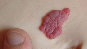
The vast majority of hemangiomas are not visible at birth and begin to appear within the first few days of life. Precursor hemangioma lesions are telangiectasias with surrounding pallor, pink macules, and bluish patch-like lesions. Pink macules and patches can mimic capillary malformations; examination of the lesion in subsequent weeks is necessary to confirm the diagnosis. The vast majority of lesions are solitary, but multiple lesions may occur in 20% of infants. Hemangiomas can appear almost anywhere on the skin and mucous membranes, but most commonly favor the head and trunk. Hemangioma sizes can vary from a few millimeters to several centimeters. Clinically, they present in 3 different forms: superficial, deep, and combined. Superficial hemangiomas are more common and clinically rise as red papules, nodules, or plaques over normal skin. Superficial hemangiomas are also known as strawberry hemangiomas or capillary hemangiomas.
Complications;
Most hemangiomas are uncomplicated and do not require intervention. Nevertheless, some lesions can cause ulceration, pain, bleeding, scar formation, and infection. Ulceration can occur in up to 10% of infantile hemangiomas and is the most common complication. Hemangiomas on the lips, anogenital area, and skin folds are more prone to ulceration. Hemangiomas in the rapid proliferation phase and in areas exposed to trauma or pressure ulcerate more frequently. Surface blanching in infants younger than three months is a harbinger of ulceration. In addition to causing pain, ulceration increases the risk of infection and results in scar formation due to the structural changes that occur. Bleeding occurs less frequently and is usually controlled by applying pressure. Bleeding: Although families of children with hemangiomas fear bleeding, bleeding is rarely profuse and can usually be stopped by applying direct pressure.
Treatment;
In hemangioma treatment, patients should be evaluated individually by considering the lesion’s size, morphology, location, the presence of potential complications, the potential to leave scars or structural deformities, the patient’s age, and the rate of growth or involution.
Treatment of Complicated Hemangiomas;
Large hemangiomas at risk of scarring or structural deformities, life-threatening hemangiomas, hemangiomas carrying a risk of functional impairment, and ulcerated hemangiomas unresponsive to standard wound care treatments can be defined as complicated hemangiomas. Propranolol: The incidental discovery in 2008 of the use of propranolol, a non-selective β-blocker, in the treatment of infantile hemangioma (upon its use in 2 children with both heart failure and hemangioma) revolutionized the management of this common tumor. Subsequently, many extensive case reports and randomized controlled trials were published; its use was approved by the FDA in 2014, and it is currently accepted as the first-line treatment for infantile hemangioma. Propranolol is thought to act via vasoconstriction, decreased expression of VEGF and bFGF, and induction of apoptosis. Thus, it is considered to inhibit growth and induce regression in infantile hemangiomas. In a randomized study conducted with 456 infants aged 1-5 months, patients were given placebo, 1 mg/kg propranolol, and 3 mg/kg propranolol for 3-6 months; at the end of the 6th month, the rate of hemangiomas that completely or nearly completely resolved was 60% in the 3 mg/kg propranolol group, compared to 4% in the placebo group. While known side effects of propranolol (hypoglycemia, hypotension, bradycardia, and bronchospasm) were reported to be observed rarely in patients, the incidence of these side effects was noted not to be significantly higher than that of placebo. In a study of 42 children aged 7 months to 10 years evaluating the use of propranolol outside the proliferation phase, using 1.5-3 mg/kg/day propranolol for 1-8 months halted hemangioma growth and demonstrated a greater increase in the involution rate in the propranolol-treated group.
Systemic Corticosteroids: Before oral β-blockers replaced systemic corticosteroids in hemangioma treatment, systemic corticosteroids were the first-line therapeutic agents for life- or function-threatening hemangiomas. Corticosteroids suppress VEGF synthesis and inhibit vasculogenesis. Systemic steroids can still be used in combination with β-blockers in cases of airway obstruction or diffuse hepatic hemangioma, or when β-blocker therapy is contraindicated. In a randomized controlled trial comparing propranolol with prednisolone, both agents showed similar efficacy, with prednisolone acting faster, but propranolol was noted to be better tolerated with a more favorable side effect profile. Although some clinicians recommend high starting doses of prednisolone such as 5-6 mg/kg/day, prednisolone is generally initiated at a dose of 2-3 mg/kg/day.
Vincristine: It is a chemotherapeutic agent used in childhood neoplasms. It inhibits microtubule formation during mitosis and induces apoptosis of tumor and endothelial cells. It is used in Kasabach-Merritt phenomenon associated with kaposiform hemangioendothelioma and tufted hemangioma. It can be used in life-threatening hemangiomas refractory to propranolol and corticosteroids. Vincristine toxicity may result in peripheral neuropathy, constipation, jaw pain, and anemia. Interferon α: It is a potent angiogenesis inhibitor. In the past, it was used as an alternative treatment agent in aggressive hemangiomas resistant to corticosteroids. Due to its potential to cause irreversible spastic diplegia and other possible complications (fever, irritability, neutropenia, liver enzyme abnormalities), it is very rarely used in the treatment of infantile hemangioma.
Surgical and Laser Treatments;
In addition to systemic, topical, and intralesional therapies, surgical and laser treatments are also among the options in hemangioma management. However, cauterization, radiotherapy, and cryotherapy are generally not recommended for infantile hemangioma treatment because they may leave more scarring than leaving the lesions untreated.
Approach in Special Circumstances;
Periorbital Hemangiomas: Superficial periocular hemangiomas can be successfully treated with topical β-blockers. Surgical excision may be an option for small, localized lesions. In sight-threatening lesions, systemic propranolol is generally recommended, whereas intralesional corticosteroid administration may be used less frequently. It should not be overlooked that intralesional corticosteroids applied around the eye may cause side effects such as eyelid necrosis, central retinal artery occlusion, and adrenal suppression.
Ulcerated Hemangiomas:
The aim in ulcerated hemangiomas is to heal the ulceration, prevent infection, and reduce pain. Superficial ulcerations can be managed with local wound care. Local wound care reduces pain and helps prevent secondary infections. Topical antibiotics, barrier creams, and non-adherent wound dressings are used in wound care. Since crusting prevents re-epithelialization, softening crusted wounds with saline solution and gently debriding crusts aids in ulcer healing. In a retrospective study of 30 children, application of topical timolol 0.5% gel once daily to focal 3 cm ulcerated hemangiomas in intertriginous areas resulted in symptom relief and partial resolution within an average of 67 days.
In the treatment of infantile hemangioma, oral propranolol has been shown to be the preferred treatment option for high-risk infantile hemangiomas, while the use of topical beta-blockers such as timolol is an important option for uncomplicated hemangiomas and has proven to be effective and safe even in cases where oral propranolol treatment cannot be administered. The contraindications and side effects of timolol are identical to those of propranolol; however, because it is applied topically, these effects occur far less frequently. It is contraindicated in patients with asthma, bradycardia, second- or third-degree atrioventricular block, or cardiogenic shock. It should also not be used on ulcerated hemangiomas or mucosa. Although some systemic absorption occurs, side effects are rare. Its use does not require monitoring. The dosage is between 1 and 4 drops per day, divided into 2 applications; the drops are applied across the entire area of the hemangioma with gentle massage. Hands should be washed with soap and water after application. Below, we see two alternative formulations published in Spain’s oldest monthly medical journal, which is the official publication of the Spanish Academy of Dermatology and Venereology (AEDV), founded in 1909.
Timolol is a highly water-soluble active ingredient. Given the characteristics of the lesions, this allows the use of hydrophilic vehicles such as gels, which are ideal and cosmetically highly acceptable for topical application.
Tuberous Sclerosis:
Rapamycin (Sirolimus) inhibits the mTOR signaling pathway, which plays an essential role in cell growth and proliferation. It can be used in complex vascular anomalies such as kaposiform hemangioendothelioma associated with Kasabach-Merritt phenomenon. There is a case report demonstrating successful rapamycin treatment in a child with PHACE(S) syndrome refractory to conventional therapies. Potential side effects include mucositis, hyperlipidemia, headache, hepatotoxicity, and neutropenia.
mTOR inhibitors have been shown to be effective and safe in the treatment of tuberous sclerosis. Various studies have demonstrated the efficacy of topical rapamycin in the treatment of facial angiofibromas characteristic of this disease. Studies have shown that a 1% concentration provides the best results, and nighttime application of the treatment is recommended.
In the formulation, due to the physical and chemical properties of the active ingredients, an oily vehicle such as petrolatum (vaseline) should be used, as its strong occlusive properties provide a greater therapeutic effect. In patients with acne or acneiform reactions, a medium-fat content emulsion can be used.
Additionally, in the treatment of ulcerations in intertriginous and moist areas such as the perineal region, the use of topical metronidazole gel combined with topical mupirocin has been found to be safe and effective. There are case series indicating that the application of becaplermin (recombinant platelet-derived growth factor) 0.01% gel accelerates healing in chronic ulcerated infantile hemangiomas. This expensive treatment has received FDA approval for the treatment of diabetic ulcers in adults. In these patients, the use of 3 or more 15 g tubes of becaplermin gel has been reported to be associated with an increase in malignancy mortality (not incidence). The use of oral propranolol can be beneficial in extensive ulcerated hemangiomas that do not heal with local wound care. In patients with ulcerated hemangiomas persisting for an average of seven weeks, the use of propranolol was reported to result in ulcer healing within an average of four weeks, and pain reduction within 15 days in most patients. Local wound care and occlusive dressings can assist with pain control. Oral acetaminophen and topical lidocaine ointment can also be effective in reducing pain; however, it should be kept in mind that lidocaine ointment may cause systemic lidocaine toxicity. The use of Eutectic mixture of local anesthetics (EMLA) in infants younger than three months should be avoided because EMLA contains prilocaine hydrochloride, which can cause infantile methemoglobinemia. Methemoglobin reductase levels are low in infants younger than three months. In ulcerated hemangiomas, 595 nm PDL laser application every 2-4 weeks combined with medical therapy and good wound care can be effective.
Conclusion;
Hemangiomas are the most common vascular tumors in infancy, and the vast majority resorbs spontaneously over time. While investigating other potential comorbid conditions in segmental hemangiomas and managing patients who develop complications are crucial, anticipating lesions likely to undergo spontaneous resolution and adopting a non-intervention strategy in consultation with patients and their relatives is just as important. In patients requiring topical or systemic treatment, knowing the benefits and side effects of the prescribed medications thoroughly is mandatory, especially since our patients are infants who cannot express their complaints and have a long life ahead of them.
Specialist Pharmacist Ahmet Nezihi Pekcan / Pekcan Pharmacy Konya
References:
1- Specialist Dr. Gökhan Şahin1, Prof. Dr. Fatma Aydın2 1 Bayburt State Hospital, Dermatology Clinic 2 Ondokuz Mayıs University Faculty of Medicine Department of Skin and Venereal Diseases Correspondence Address: Dr. Fatma Aydın, Ondokuz Mayıs University Faculty of Medicine Department of Dermatology 55139 Kurupelit Samsun Turkey E-mail: [email protected]
2- Actas Dermo-Sifiliográficas The Most Useful Pharmaceutical Formulations (Individualized Medications) in Pediatric Dermatology: A Review E. Abarca Lachén,a,b,∗ P. Hernando Martínez,a,b Y. Gilaberte Calzadac a Facultad de Ciencias de la Salud, Universidad San Jorge, Villanueva de Gállego, Zaragoza, Spain b Sociedad Espanola ˜ del Medicamento Individualizado (LASEMI), Spain c Servicio de Dermatología, Hospital Universitario Miguel Servet, IIS Aragón, Zaragoza, Spain Received 18 August 2020; accepted 9 November 2020 Available online 2 February 2021